Ulcerative colitis (UC) is a chronic inflammatory bowel disease (IBD) characterized by inflammation in the lining of the colon and rectum. Diagnosing UC accurately is crucial for effective management and treatment. The process involves a combination of clinical evaluation, laboratory tests, and diagnostic imaging to confirm the presence of the disease and assess its severity. This article outlines how ulcerative colitis is diagnosed along with the standard steps and methods that are used.
Initial Assessment
The diagnosis of ulcerative colitis typically begins with a detailed medical history and a physical examination. Patients often report symptoms such as prolonged diarrhea, blood in the stool, abdominal pain, and significant weight loss. Doctors will inquire about the duration and severity of these symptoms, family history of IBD, and any related autoimmune conditions. During the physical exam, physicians may check for tenderness in the abdomen, signs of anemia, and other indicators of inflammation.
Laboratory Tests
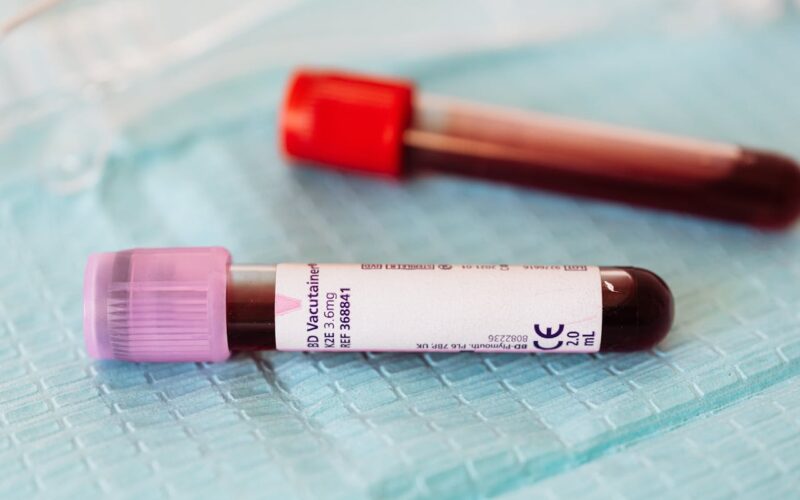
In the diagnostic process for ulcerative colitis, blood tests play a crucial role in assessing the overall health and presence of inflammation. Specifically, these tests often include a complete blood count (CBC) to check for anemia, which can occur from chronic or intense bleeding in the colon, and to measure white blood cell counts. Elevated white blood cell counts are a common indicator of inflammation or infection somewhere in the body, including in the gastrointestinal tract.
In addition to blood tests, doctors frequently utilize stool tests as part of the diagnostic process. Stool tests are valuable for several reasons when diagnosing ulcerative colitis. Firstly, they can detect the presence of white blood cells in the stool, which is another sign of inflammation. The absence of these cells can sometimes help rule out inflammatory causes of gastrointestinal symptoms.
Moreover, stool tests are essential for excluding infections that can mimic the symptoms of ulcerative colitis. For instance, an infection with Clostridioides difficile (C. diff), a bacteria that causes symptoms such as diarrhea, fever, and abdominal pain, must be ruled out. This is particularly important as C. diff can cause severe complications and is treated differently than ulcerative colitis. Doctors might also look for other pathogens such as parasites or bacteria like Salmonella and Shigella, which can produce similar gastrointestinal symptoms. Confirming that these pathogens are not the cause of the symptoms ensures that treatments specific to ulcerative colitis can be initiated without delay.
Endoscopic Procedures
Endoscopy is the most definitive method for diagnosing ulcerative colitis. The two primary types of endoscopic procedures used are:
- Colonoscopy: This is the most comprehensive tool for examining the entire colon and rectum. During a colonoscopy, a long, flexible tube with a camera on the end (colonoscope) is inserted through the anus to view the entire colon. The doctor can observe the pattern of inflammation and take tissue samples (biopsies) for further pathological examination.
- Flexible Sigmoidoscopy: Less extensive than a colonoscopy, this procedure involves examining only the lower part of the colon (sigmoid) with a flexible viewing tube. It is used when the symptoms are primarily located in the lower colon or if there are risks that make a full colonoscopy inadvisable.
Imaging Studies
In some cases, additional imaging studies might be required to diagnose complications or severe cases of UC. These might include:
- X-rays: Although not routinely used to diagnose UC itself, X-rays can be helpful in identifying complications such as a perforated colon.
- CT scans: These are often used to get a more detailed view of the colon and surrounding tissues, especially if complications such as abscesses or fistulas are suspected.
- MRI: An MRI may be employed to assess the severity of inflammation or to check for complications in the pelvic area.
Differential Diagnosis
Differentiating ulcerative colitis from other similar conditions is crucial for effective treatment, and it involves examining several key aspects:
Location of Inflammation: Ulcerative Colitis is typically confined to the colon and rectum and presents a continuous pattern of inflammation starting from the rectum and extending upwards. In contrast, Crohn’s disease can affect any part of the gastrointestinal tract from mouth to anus and shows patchy, non-continuous inflammation.
Depth of Inflammation: The inflammation in UC affects only the innermost lining of the colon, making it relatively superficial. Crohn’s disease, however, features transmural inflammation, which can penetrate multiple or all layers of the bowel wall. This deep inflammation can lead to complications such as fistulas and strictures, which are less common in UC.
Pattern of Symptoms: Both ulcerative colitis and Crohn’s disease can cause symptoms like diarrhea, rectal bleeding, and abdominal pain. However, UC typically results in a constant urge to defecate due to inflammation concentrated in the lower colon and rectum. Crohn’s disease symptoms may include more generalized abdominal pain and significant weight loss due to broader areas of the digestive tract being affected.
By carefully assessing these factors—location, depth, and symptom patterns—healthcare providers can accurately diagnose ulcerative colitis and differentiate it from conditions such as Crohn’s disease and infectious colitis. This precision is vital to ensure that patients receive the most appropriate and effective management strategies tailored to their specific condition.
Conclusion: Key Steps in Diagnosing Ulcerative Colitis
By carefully assessing factors such as the location and depth of inflammation, as well as the specific patterns of symptoms, healthcare providers can accurately diagnose ulcerative colitis. This precision is crucial for ensuring that patients receive the most appropriate and effective treatment strategies tailored to their specific condition. Accurate diagnosis not only helps in differentiating UC from similar conditions like Crohn’s disease and infectious colitis but also guides the management and improves the overall quality of life for those affected.


